قطعه بندی تصاویر رادیوگرافی پانورامیک دندانی با استفاده از روش های یادگیری عمیق مبتنی بر ساختار U-Net
کد: G-1957
نویسندگان: Nastaran Moeinfar ℗, Seyyede Zohreh Seyyedsalehi *
زمان بندی: زمان بندی نشده!
برچسب: پردازش سیگنال های پزشکی
دانلود: دانلود پوستر
خلاصه مقاله:
خلاصه مقاله
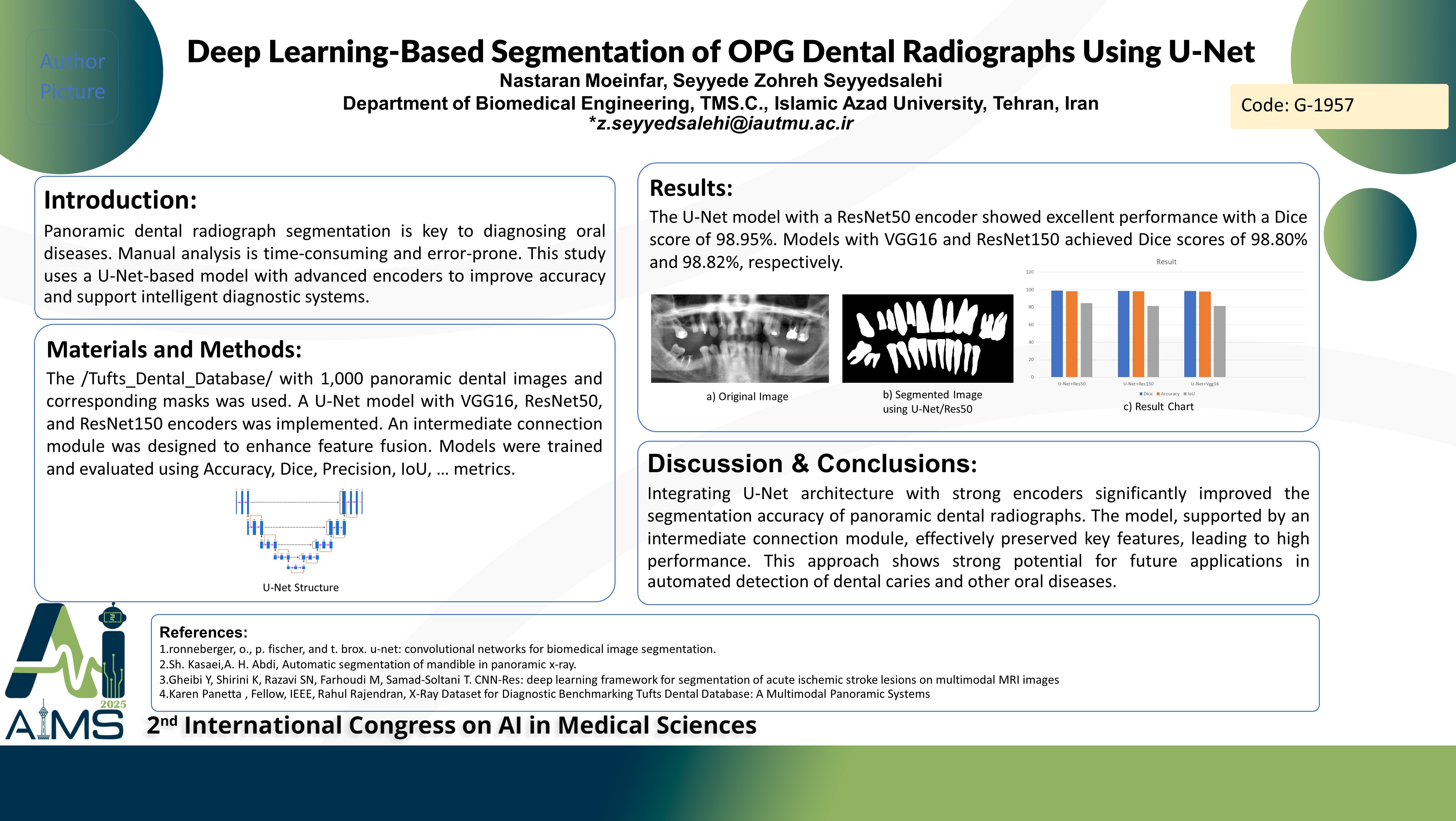
Manual interpretation of dental radiographs is time-consuming and prone to error. Deep learning offers an efficient solution by automating the segmentation process, enabling faster and more accurate diagnoses. Various segmentation methods have been explored in previous studies, though many failed to deliver satisfactory accuracy. This research aims to improve segmentation performance by utilizing an enhanced U-Net architecture integrated with advanced encoders. Background and aims: Artificial intelligence (AI), particularly in the realm of computer vision, plays a vital role in enhancing both accuracy and efficiency across various domains. Image segmentation, a core technique within medical image analysis, is the focus of this study. The goal is to develop a deep learning-based model tailored to the segmentation of panoramic dental radiographs. Method: The study employed the Tufts_Dental_Database, which includes 1,000 panoramic dental radiographs paired with their corresponding masks. A U-Net architecture was selected for its encoder-decoder framework and effective use of skip connections. To improve feature fusion and retention, a custom intermediate connection module was incorporated into the network. Three encoder backbones VGG16, ResNet50, and ResNet150 were integrated separately into the U-Net framework for comparison. All models were trained with a batch size of 1 over 50 epochs. Training beyond50epochs showed no meaningful improvement. Evaluation was conducted using standard metrics including Accuracy, Precision, Recall, Intersection over Union (IoU), and the Dice coefficient. Results: The Dice coefficient evaluation indicated strong segmentation performance across all models. The ResNet50-based U-Net achieved the highest Dice score of 98.95%, followed by models incorporating VGG16 (98.80%) and ResNet150 (98.82%). These findings underscore the effectiveness of the U-Net structure and the contribution of the intermediate connection module in enhancing segmentation accuracy. Conclusion: Integrating deep encoders with a refined U-Net architecture enables precise segmentation of dental radiographs. The network’s design particularly the intermediate connection module facilitates robust feature extraction and fusion. This model lays a solid foundation for intelligent diagnostic tools and may be adapted for the detection and segmentation of dental caries and other oral conditions.
کلمات کلیدی
Deep Learning, Segmentation, U-Net, Tooth Diagnosis