The Role of Radiomics in Multiple Sclerosis Imaging
کد: G-1642
نویسندگان: Kimia Kazemzadeh * ℗
زمان بندی: زمان بندی نشده!
برچسب: پردازش سیگنال های پزشکی
دانلود: دانلود پوستر
خلاصه مقاله:
خلاصه مقاله
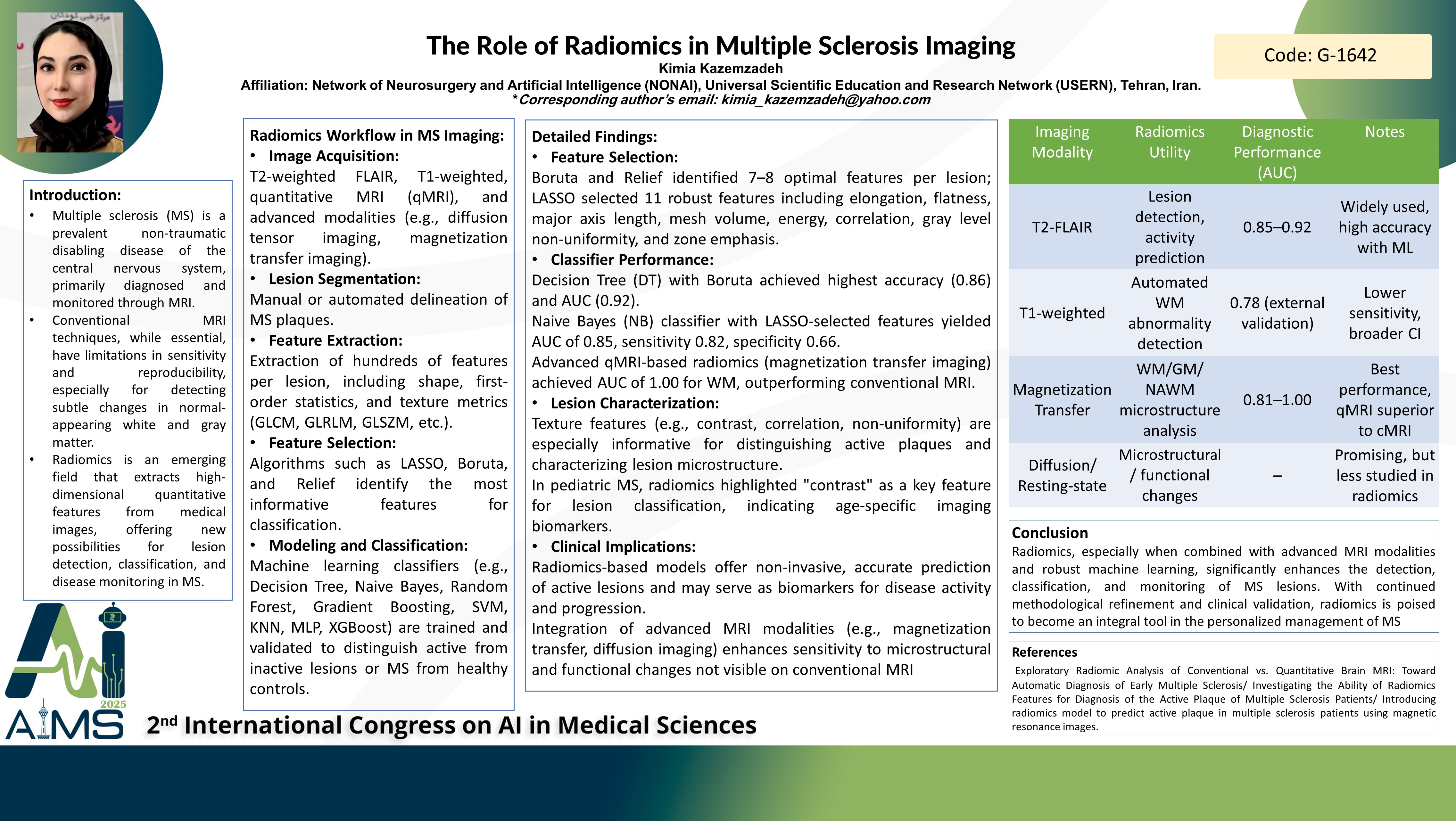
Background and Aim: Multiple sclerosis (MS) is a common non-traumatic disabling disease frequently diagnosed through magnetic resonance imaging (MRI). Radiomics, an emerging field that extracts quantitative features from medical images, has shown promise in improving the diagnostic accuracy of active MS plaques and predicting disease progression. This review aims to evaluate the role of radiomics in MS imaging, focusing on its potential to enhance lesion detection, classification, and disease monitoring. Method: This narrative review synthesizes findings from various studies investigating radiomic applications in MS imaging. It covers the use of radiomic features extracted from T2-weighted Fluid Attenuated Inversion Recovery (FLAIR) images, the application of feature selection algorithms such as LASSO, Boruta, and Relief, and the employment of machine learning classifiers including K-Nearest Neighbors, Random Forest, Naive Bayes, Decision Trees, and Extreme Gradient Boosting. The review also explores radiomic analyses incorporating advanced MRI techniques like diffusion tensor imaging and resting-state functional MRI to assess microstructural and functional brain changes in MS. Result: Radiomic features derived from T2-FLAIR images have been effective in predicting active MS plaques, with the Naive Bayes classifier achieving an area under the curve (AUC) of 0.85, demonstrating non-invasive prediction capability. Feature selection combined with classifiers such as Boruta and Decision Trees yielded an average accuracy of 0.86 and an AUC of 0.92, highlighting the critical role of algorithm choice in diagnostic performance. In pediatric MS patients, radiomic models based on T2-FLAIR images identified specific features, particularly "contrast," as significant for lesion classification, suggesting enhanced lesion characterization in younger populations. Incorporating advanced MRI modalities into radiomic workflows provided further insights into MS-related microstructural and functional brain alterations. Despite these advances, challenges remain, including the need for standardization of radiomic methodologies, validation in larger cohorts, and integration into clinical decision-making to fully realize radiomics' clinical utility in MS. Conclusion: Radiomics represents a promising advancement in MS imaging by improving the detection and characterization of active lesions and offering potential for disease progression prediction. Continued refinement, validation, and clinical integration of radiomic approaches are essential to harness their full potential in enhancing MS diagnosis and management
کلمات کلیدی
Multiple sclerosis, Radiomics, Imaging