emerging role of digital pathology to advance cancer immunotherapy: a systematic review
Code: G-1572
Authors: Shahin Akhoundi * ℗, Samad Nadri, Seyedeh Negin Mousavi, Amirhossein Shojaei
Schedule: Not Scheduled!
Tag: Cancer Diagnosis & Treatment
Download: Download Poster
Abstract:
Abstract
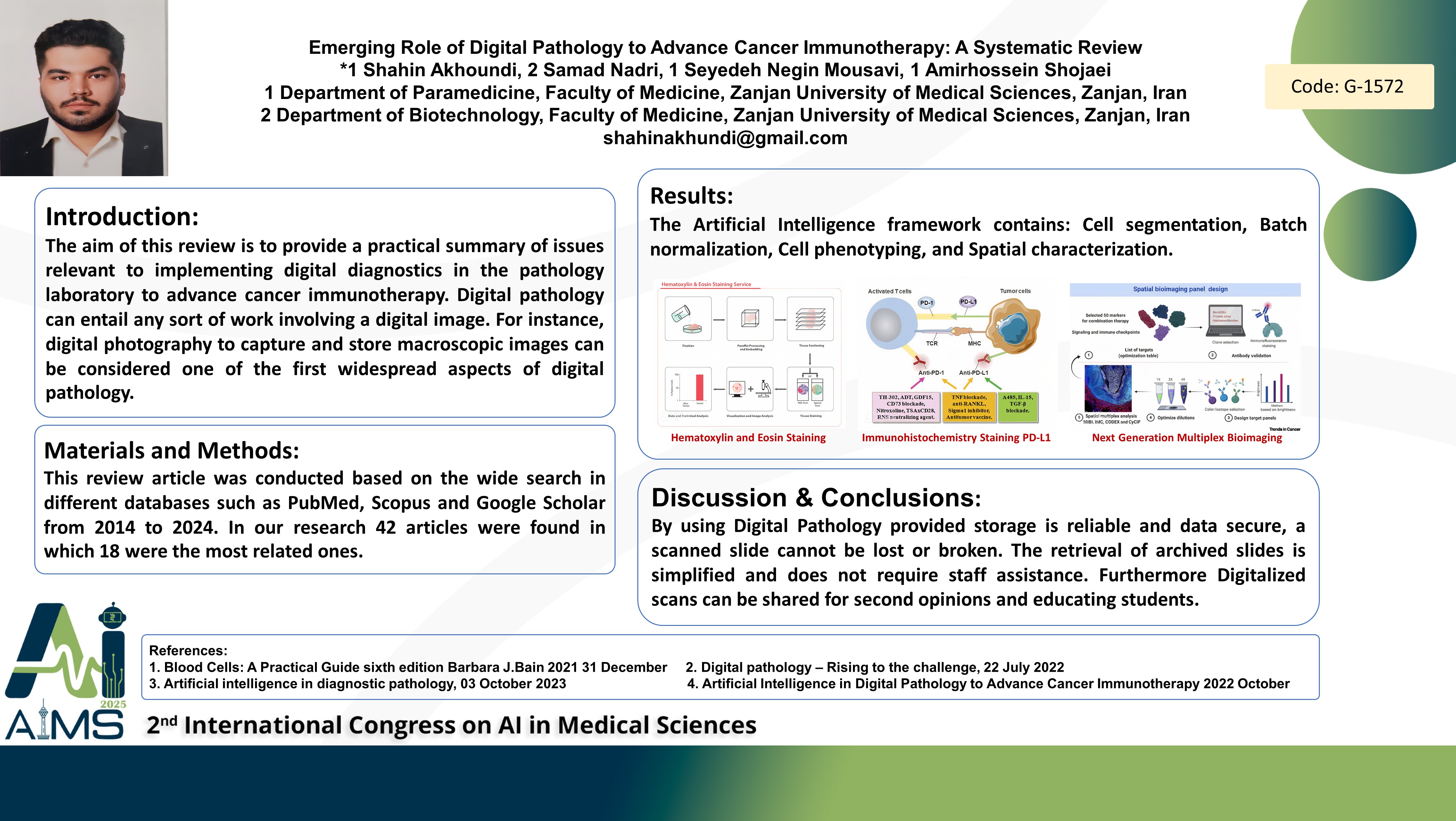
Background and aims: The aim of this review is to provide a practical summary of issues relevant to implementing digital diagnostics in the pathology laboratory to advance cancer immunotherapy. Digital pathology can entail any sort of work involving a digital image. For instance, digital photography to capture and store macroscopic images can be considered one of the first widespread aspects of digital pathology. With the rapid development of hardware and software for whole slide imaging, particularly since the Food and Drug Administration approved this system to be used for primary diagnosis, digital pathology has gained substantial momentum in scientific research. Computer-aided diagnosis is embedded into the pathology flow powered by the digital pathology framework. Method: This review article was conducted based on the wide search in different databases such as PubMed, Scopus and Google Scholar from 2014 to 2024. In our research 42 articles were found in which 18 were the most related ones. Results: The Artificial Intelligence framework contains: Cell segmentation, Batch normalization, Cell phenotyping, and Spatial characterization. Routine Hematoxylin and Eosin Staining: H&E is used to reveal the nuclei morphology and from which tumor-infiltrating lymphocytes have demonstrated strong clinical values related to immunotherapy in various tumor types. With Artificial intelligence we can access to the cells more quickly and more confident. Immunohistochemistry Staining: PD-L1, is clinically used to stratify immune-checkpoint inhibitors based on therapies in lung and other cancer types. There are studies that developed AI-enabled automatic PD-L1 scoring systems based on assays in breast, head and neck cancer. Next Generation Multiplex Bioimaging: It can profile proteins in situ, enabling the systematic study of cellular compositions and interactions. The multiplex imaging with big data imposes a challenge for pathologists to directly evaluate these slides, where AI-powered tools are being developed to characterize the tumor microenvironment. Conclusion: By using Digital Pathology provided storage is reliable and data secure, a scanned slide cannot be lost or broken. The retrieval of archived slides is simplified and does not require staff assistance. Furtheremore Digitalized scans can be shared for second opinions and educating students.
Keywords
Digital Pathology, Artificial Intelligence, Cancer, Immunotherapy