بررسی نحوه خون رسانی به بافت مغز با استفاده از تصاویر SPECTمبتنی بر هوش مصنوعی
کد: G-1436
نویسندگان: Pariyan Monshi Karimi *, Mahdi Baghbani ℗, Mohammad-Reza S. Noorani
زمان بندی: زمان بندی نشده!
برچسب: رباتیک در جراحی و مراقبت سلامت
دانلود: دانلود پوستر
خلاصه مقاله:
خلاصه مقاله
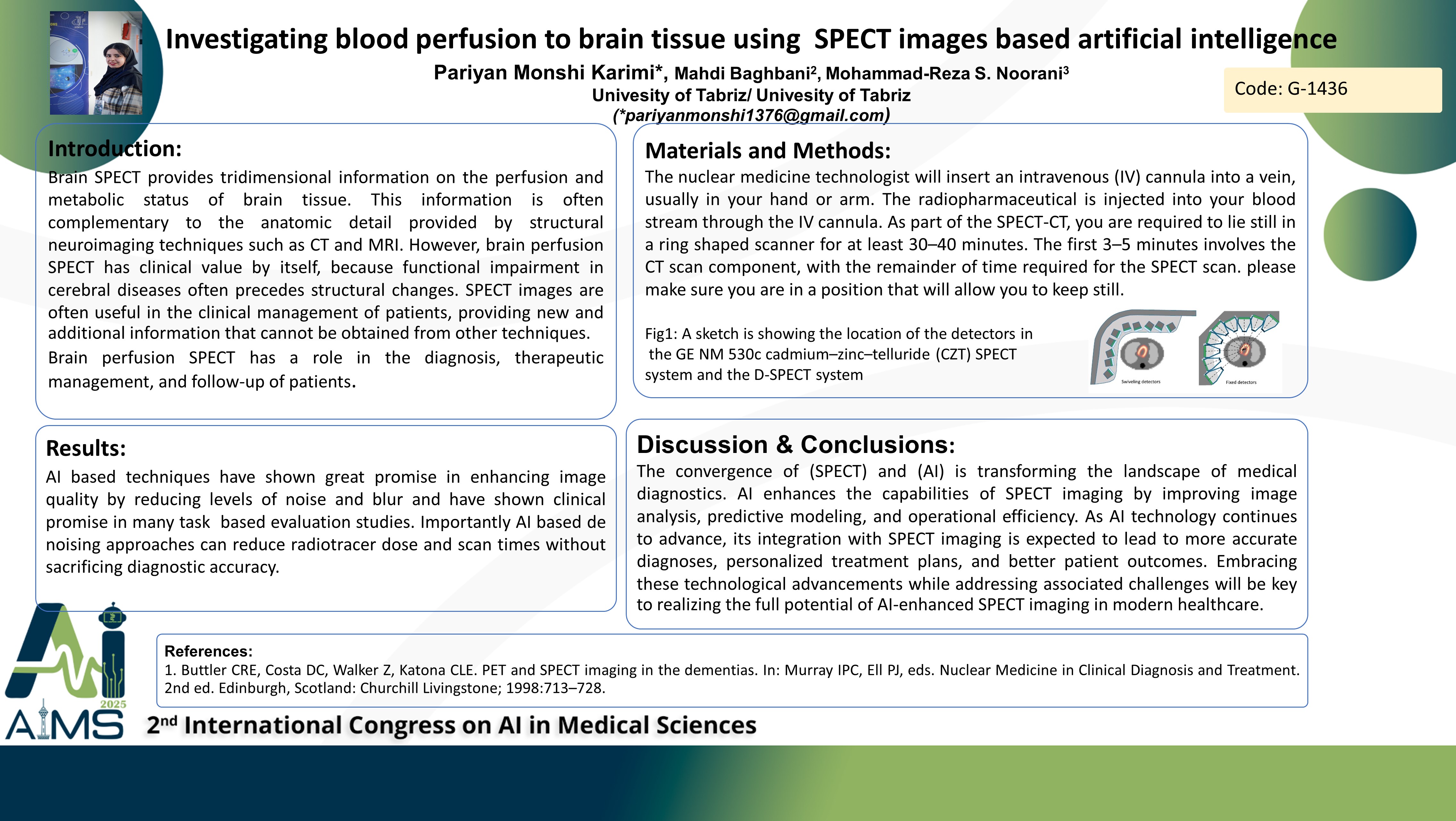
Single-photon emission computed tomography (SPECT) is a nuclear imaging modality frequently used in diagnostic medicine. At its most basic level, SPECT produces a three-dimensional image of the distribution of a radioactive tracer injected into the blood stream and subsequently taken up by certain tissues. Brain Perfusion SPECT scan is a type of nuclear medicine imaging test that measures blood perfusion in the brain, investigation of sensorial, motor, and cognitive activities and the central effects of central nervous system, drugs in both the normal and the abnormal brain. Recently, many artificial intelligence (AI) algorithms have been proposed to compensate for such deficiencies. Studies include denoising and resolution improvement for SPECT images. Background and aims: Obtaining three-dimensional information about brain perfusion and metabolic status, pathological status of the patient. Diagnosing cerebral perfusion abnormalities with the help of tracer absorption rate. Method: the nuclear medicine technologist will insert an intravenous (IV) cannula into a vein, usually in your hand or arm. The radiopharmaceutical is injected into your blood stream through the IV cannula. You will then be required to lie on a bed while detectors or cameras obtain the scan images. As part of the SPECT-CT, you are required to lie still in a ring shaped scanner for at least 30–40 minutes. The first 3–5 minutes involves the CT scan component, with the remainder of time required for the SPECT scan. It is very important that you remain still for the entire duration of the two scans, so that the SPECT and CT can be accurately combined. Results: AI based techniques have shown great promise in enhancing image quality by reducing levels of noise and blur and have shown clinical promise in many task based evaluation studies. Importantly, many studies have suggested that AI based de noising approaches can reduce radiotracer dose and scan times without sacrificing diagnostic accuracy. Conclusion: The convergence of SPECT and AI is transforming the landscape of medical diagnostics. AI enhances the capabilities of SPECT imaging by improving image analysis, predictive modeling, and operational efficiency.
کلمات کلیدی
Artificial Intelligence, Brain Perfusion, SPECT